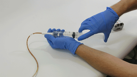
fbpx

Acidic Fluids May Coagulate or Thicken Clogs – Tuesday Tube Facts

Did you know…?

The use of acidic liquids to clear a clogged feeding tube may make the problem worse.[1,2]

Enteral feeding proteins coagulate or thicken in an acidic environment.[1,2]

References:

  1. Dandeles LM, Lodolce AE. Efficacy of agents to prevent and treat enteral feeding tube clogs. Ann Pharmacother. 2011;45:676-680.
  2. Williams NT.Medication administration through enteral feeding tubes. Am J Health-Syst Pharm. 2008;65:2347-2357.

Fluoroscopy EAD Replacement Costs – Tuesday Tube Facts

Did you know…?

Using fluoroscopy to replace a clogged feeding tube can cost as much as $1,000.[1]

In addition, replacement often exposes the patient to additional radiation.[2]

References:

  1. Fisher C, Blalock B. Clogged feeding tubes: a clinician’s thorn. Pract Gastroenterol. 2014;38:16-22.
  2. Garrison, C. M. Enteral Feeding Tube Clogging: What Are the Causes and What Are the Answers? A Bench Top Analysis. Nutrition in Clinical Practice. 2018. 33(1):147-150.

Clogging: A Frequent Complication in EADs – Tuesday Tube Facts

Did you know…?

Clogging is one of the most frequent mechanical complications of feeding tubes.[1,3]

Reported clogging rates vary from 9 – 35%.[1-7] With clogging of nasogastric (NG) feeding tubes considered under-reported, the actual rate may be much higher.[3]

References:

  1. Ireton-Jones C, DeLegge M. Handbook of Home Nutrition Support. Sudburry, MA: Jones and Bartlett; 2007.
  2. Beyer PL, Matarese LE, Gottschlich MM. Complications of enteral nutrition. 1998.
  3. Blumenstein I, Shastri YM, Stein J. Gastroenteric tube feeding: techniques, problems and solutions. World J Gastroenterol. 2014;20(26):8505-8524.
  4. Pancorbo-Hidalgo PL, García-Fernandez FP, Ramírez-Pérez C. Complications associated with enteral nutrition by nasogastric tube in an internal medicine unit. J Clin Nurs. 2001;10(4):482-490.
  5. Marcuard SP, Stegall KS. Unclogging feeding tubes with pancreatic enzyme. JPEN J Parenter Enteral Nutr. 1990;14(2):198-200.
  6. Frizzi JD, Ray PD, Raff JB. Enteral nutrition by a forward surgical team in Afghanistan. South Med J. 2005;98(3):273-278.
  7. Bourgault AM, Heyland DK, Drover JW, Keefe L, Newman P, Day AG. Prophylactic pancreatic enzymes to reduce feeding tube occlusions. Nutr Clin Pract. 2003;18(5):398-401.

Syringe Force on Clogged Feeding Tubes

Syringe Force on Clogged Feeding Tubes

Many people think feeding tubes are merely used to deliver nutrition (i.e., feeding formula), however, they deliver life-sustaining medications too – it’s no wonder they tend to clog. At Actuated Medical, Inc., we developed the TubeClear System to keep enteral therapy (medication, nutrition and hydration) being delivered as prescribed.


i

Article Summary

  • Forces required to do air flushes via enteral syringe in feeding tubes with no clog, 1-layer, and 2-layers of feeding formula mixtures were measured.
  • Feeding tube clogs, or material build-up on the walls (i.e., 1-layer clogs), may begin forming before becoming noticeably difficult to flush.
  • 2-layer clogs required a force that was nearly 5.5 times of the “No Clog” feeding tubes, which was noticeable when flushed by hand.
  • The TubeClear System should be used when caregivers notice higher forces during air or water flushes to maintain full patency of feeding tubes.

We have demonstrated that compared to flushing with water alone, the TubeClear System can remove residue “sludge” from feeding tube interior walls with greater efficacy.[1] However, the question remains – How does one know when material build-up is adhering to the interior feeding tube wall if fluids can still be introduced into the feeding tube?

For patients using feeding pumps, most pump alarms will sound an alert when the flow rate falls below the predesignated flow rate.[2] However, for those using gravity or syringe feedings, clinicians flush the feeding tube with a syringe to confirm patency (open or unobstructed). These water flushes may require increased force if materials are building up on the inner tube walls. In cases of fully clogged feeding tubes, the water may blow backward toward the clinician due to increased syringe resistance. Note that using excessive force on a syringe (often called power flushing) is generally not recommended as it may cause a rupture to the patient’s feeding tube.[3]

Measuring Flushing Force

Our engineers investigated the amount of force (Newtons [N]) required to be applied to a syringe plunger when attempting to introduce air into non-clogged and partially clogged feeding tubes. In vitro produced clogs were created with a mixture of feeding formula and fiber within 14Fr NG feeding tubes (Cardinal Health, Dublin, OH). Three (3) different feeding tube conditions were tested, N=15 trials, with n=5 trials/feeding tube condition: 1) No clog (lacking all material), 2) 1-layer of clog mixture on feeding tube walls, and 3) 2-layers of clog mixture on feeding tube walls (see Figure 2). To create the single layer, the clog mixture was introduced into feeding tubes (N=10) and dried at 55°C for three (3) hours.  To create the two-layer clog buildup, a second clog layer was introduced into a subset of the same feeding tubes (N=5) and dried at 55°C for three (3) hours. To measure the force, a 60-cc syringe was filled with air to the 50-cc mark, and the plunger was depressed to the 10-cc mark using a Shimpo model 100XY force gauge (ELECTROMATIC Equipment Co., Inc., Cedarhurst, NY) (see Figure 1). A n = 5 trials were performed for each feeding tube condition.

Figure 1: Testing set-up.

Figure 2: A rendering of the cross-section of the clog types. The grey circle represents the feeding tube wall, and clog material is represented in shades of orange.

The peak force (measured in Newtons [N]) required to push air via the syringe through a non-clogged feeding tube averaged 7.6±0.1N, compared to 14.3±0.5N and 41.0±1.7N, for the one and two layered partially clogged feeding tubes, respectively (see Figure 3).

While these measurements demonstrate an increasing amount of required force, what does that feel like in practice?

Figure 3: Syringe Force testing summary. 
Blue Bar= No clog, Orange Bar= Single layer of clog mixture, Gray Bar= Double layer of clog mixture. n=5 trials/performed for each feeding tube condition. Error bars= standard error of the mean. Two-way ANOVA, Tukey’s Multiple comparison test, different variables =p<0.01.

Feeling the Force

There are many publications discussing preventative means to feeding tube clog formation and multiple ways to remove clogs;[4-5] however, the question as to how one can determine if a feeding tube is fully patent does not appear to have been addressed.

Some feeding tube clogs appear to form quickly, while others occur gradually over time, leading clinicians and care takers to be uncertain to the state of patency of a feeding tube at any particular time. We aimed to quantify the amount of required force to introduce air into feeding tubes with the various levels of clogging- with the end goal to correlate those values with a subjective “feel,” as such, initial tests were conducted without a force gauge. Videos demonstrate how easily (or not) air could be introduced into feeding tubes of the various conditions.

A

B

C

Figure 4: Syringe resistance against each feeding tube condition. A) No clog. B) 1 layer. C) 2 layers (note: looping GIF animation may look like the plunger “snaps” back, but this is the beginning of the animation).

As expected, the feeding tube without a clog (No Clog) could be flushed with air without experiencing any major resistance. However, the feeding tube with a single layer of clog mixture appears to flush almost as easily as the “No Clog” feeding tube, but the required force as measured by the force gauge was nearly double. This finding suggests that feeding tube clogs, or material build-up on the walls, may begin forming before becoming noticeable to clinicians or caretakers. Further research is required to elucidate this finding.

Attempts to flush air into feeding tubes with a double layer of clog mixture proved the most difficult, requiring additional force as the syringe plunger pushed backwards towards the Operator (see Figure 4c) due to the partial blockage. These simulated clogs required a force that was nearly 5.5 times that of the “No Clog” feeding tubes.

From Bench to Bedside

Using the TubeClear System proactively can remove material build-up from feeding tube inner walls enabling enteral therapy (i.e., medication, nutrition and hydration) to be delivered as prescribed.[1] Based on the results presented here, we recommend using the TubeClear System on a patient’s feeding tube as soon as the clinician notices a change in the required pressure to administer fluid or air during routine syringe flushing, as this may be indicative of material build-up on the inner walls of the tube.

How often do you notice a change in the required force during routine flushes? Let us know! Find us on Twitter and Facebook or send us an email at info@tubeclear.com.

References:

  1. Actuated Medical, Inc. Internal Test Report Doc. No. 1100791569-001.
  2. Cardinal Health, Dublin, OH. https://www.cardinalhealth.com/content/dam/corp/web/documents/patient-recovery/Literature/kangaroo-connect-enteral-feeding-pump-operator-manual.pdf. Accessed 9/27/19.
  3. Richards, S., Walsh, K. (2016), Feeding Tube Irrigation: Performing. Nursing Practice and Skill. https://www.ebscohost.com/assets-sample-content/Feeding_Tube_Irrigation_Performing_-_NSP.pdf. Accessed 9/27/19.
  4. Boullata, J. I., Carrera, A. L., Harvey, L. , Escuro, A. A., Hudson, L. , Mays, A. , McGinnis, C. , Wessel, J. J., Bajpai, S. , Beebe, M. L., Kinn, T. J., Klang, M. G., Lord, L. , Martin, K. , Pompeii‐Wolfe, C. , Sullivan, J. , Wood, A. , Malone, A. , Guenter, P. and , (2017), ASPEN Safe Practices for Enteral Nutrition Therapy. Journal of Parenteral and Enteral Nutrition, 41: 15-103 0148607116673053. doi:1177/0148607116673053.
  5. Lord, L.M. (2018), Enteral Access Devices: Types, Function, Care, and Challenges. Nutrition in Clinical Practice, 33(1): 16-38. doi: 10.1002/ncp.10019.
Starting EN More than 3 Days After ICU Admission – Tuesday Tube Facts

Starting EN More than 3 Days After ICU Admission – Tuesday Tube Facts

Starting enteral nutrition more than 3 days after ICU admission was associated with increased length of stay.

Critically ill patients should be started on enteral tube feeds within 48 hours of intubation whenever possible.

1. Bourgault, A.M., Ipe, L., Weaver, J., Swartz, S., O’Dea, P.J. Development of Evidence-Based Guidelines and Critical Care Nurses ’ Knowledge of Enteral Feeding. Critical Care Nurse, 2007; 27(4):17-29.

2. Fremont, R. D., Rice, T.W. How soon should we start interventional feeding in the ICU? Curr Opin Gastroenterol. 2014 March; 30(2): 178–181.
Ask ChatGPT
Set ChatGPT API key
Find your Secret API key in your ChatGPT User settings and paste it here to connect ChatGPT with your Tutor LMS website.